


Gonarthrosis of the knee joint is the most common localization of a degenerative-dystrophic disease, characterized by gradual destruction of the cartilage with subsequent changes in the articular surfaces, which is accompanied by pain and reduced mobility.
The disease is more likely to affect women over 40 years of age, especially those who are overweight and have varicose veins in the lower extremities.
The knee joint consists of three compartments:
- medial tibiofemoral;
- lateral tibiofemoral;
- suprapatellar-femoral.
These compartments can be affected individually or in any combination by deforming arthrosis (DOA). 75% of all cases of gonarthrosis are the destruction of the medial tibiofemoral compartment (during movements it experiences a load that exceeds body weight by 2-3 times).
In young patients, more often only one joint is destroyed - the right or left (right-sided or left-sided gonarthrosis).
Causes of DOA of the knee joint
Several factors can be simultaneously involved in the development of degenerative cartilage changes:
- mechanical overload of the knee joint (some specialties, sports) with microtraumatization of the cartilage;
- Consequences of injuries, surgical interventions (meniscectomy);
- inflammatory diseases of the knee (arthritis);
- anatomical inconsistencies of the articular surfaces (dysplasia);
- violation of statics (flat feet, curvature of the spine);
- chronic hemarthrosis (blood accumulation in the synovial cavity);
- metabolic pathology (gout, hemochromatosis, chondrocalcinosis);
- overweight;
- violations of the blood supply to the bone;
- osteodystrophy (Paget's disease);
- neurological diseases, loss of feeling in the limbs;
- endocrine disorders (acromegaly, diabetes mellitus, amenorrhea, hyperparathyroidism);
- genetic predisposition (generalized forms of arthrosis);
- Violation of the synthesis of collagen type II.
But in 40% of cases it is impossible to establish the actual cause of the disease (primary arthrosis).
Pathogenesis of gonarthrosis
first phase
In the initial stage of the disease, the processes of cartilage metabolism are disturbed. The synthesis and quality of the main structural unit of cartilage tissue, proteoglycans, which are responsible for the stability of the structure of the collagen network, are reduced.
As a result, chondroitin sulfate, keratin, hyaluronic acid are washed out of the mesh and structurally defective proteoglycans can no longer hold water. It is absorbed into collagen, the swollen fibers of which lead to a reduction in the cartilage's resistance to stress.
Pro-inflammatory substances accumulate in the synovial cavity, under the influence of which the cartilage is destroyed even faster. Fibrosis of the joint capsule develops. The changed composition of the synovial fluid makes it more difficult to supply the cartilage with nutrients and impairs the gliding of the articular surfaces during movement.
progression of the pathology
In the future, the cartilage gradually thins, becomes rough, cracks form throughout its thickness. The epiphyses of the bones experience an increased load, which provokes the development of osteosclerosis and compensatory proliferation of bone tissue (osteophytes).
This response of the body is aimed at increasing the area of the joint surfaces and redistributing the load. But the presence of osteophytes increases discomfort, deformity and further limits the mobility of the limb.
Microfractures are formed in the thickness of the bone, injuring the vessels and leading to intraosseous hypertension. In the last stage of arthrosis, the articular surfaces are completely exposed, deformed, the movements of the limbs are severely limited.
Symptoms of gonarthrosis of the knee joint
Arthrosis of the knee joint is characterized by a chronic, slowly progressing course (months, years). The clinic is growing gradually, without pronounced exacerbations. The patient cannot remember exactly when the first symptoms appeared.
Clinical manifestations of gonarthrosis:
- Pains. At first diffuse, short (with prolonged standing, climbing stairs) and with progressive arthrosis, the pain becomes local (front and inner surface of the knee), its intensity increases;
- local sensitivity on palpation. Usually on the inside of the knee along the edge of the joint space;
- Crunch. In stage I it can be inaudible, in stage II-III it accompanies all movements;
- Increase in volume, deformation of the knee. As a result of the weakening of the collateral ligaments, a person develops an O-shaped configuration of the limbs (it is clearly visible even in the photo);
- restriction of mobility. Initially there are difficulties with bending the knee, later - with stretching.
Causes of pain in DOA:
- mechanical friction of damaged joint surfaces;
- increased intraosseous pressure, venous congestion;
- accession of synovitis;
- Changes in periarticular tissues (stretching of the capsule, ligaments, tendons);
- thickening of the periosteum;
- phenomena of dystrophy in the adjacent muscles;
- fibromyalgia;
- compression of nerve endings.
In contrast to coxarthrosis, the symptoms of DOA of the knee can resolve spontaneously.
Clinical manifestations of gonarthrosis depending on the stage:
| Characteristics | I direct | Stage II | III. stage |
|---|---|---|---|
| Pains | Short, occurs more often when the knee is extended (prolonged standing, climbing stairs) | Moderate, disappears after a night's rest | Pronounced, also disturbing at night |
| mobility impairment | Not visible | There is a limitation in extension, slight lameness | Persistent flexion-extensor contractures, lameness |
| crunch | no | Palpable on palpation during movement | remote crunch |
| deformation | Absence | Slight forward deviation of the axis of the extremities, muscle wasting | Valgus or varus deformity. The joint is unstable, atrophy of the thigh muscles |
| X-ray picture | Slight narrowing of the joint space, first signs of subchondral osteosclerosis | The joint space is narrowed by 50% or more, osteophytes appear | Almost complete absence of the joint space, marked deformation and sclerosis of the articular surfaces, subchondral bone necrosis, osteoporosis |
A common complication of arthrosis of the knee joint is secondary reactive synovitis, which is characterized by the following symptoms:
- increasing pain;
- swelling;
- effusion into the synovial cavity;
- increase in skin temperature.
Less frequent and more dangerous complications are: blockage of the joint, osteonecrosis of the femoral condyle, subluxation of the patella, spontaneous hemarthrosis.
Diagnosis of a DOA of the knee joint
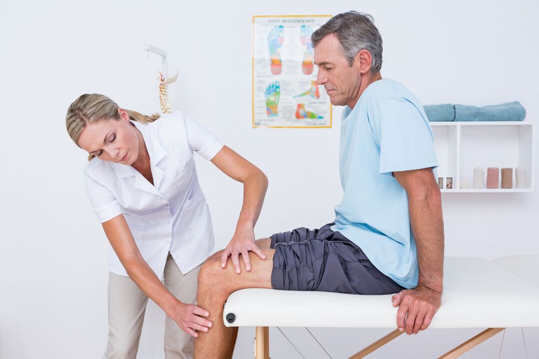
The diagnosis of gonarthrosis is based on the patient's characteristic complaints, the changes detected during the examination and the results of additional tests.
To confirm arthrosis is prescribed:
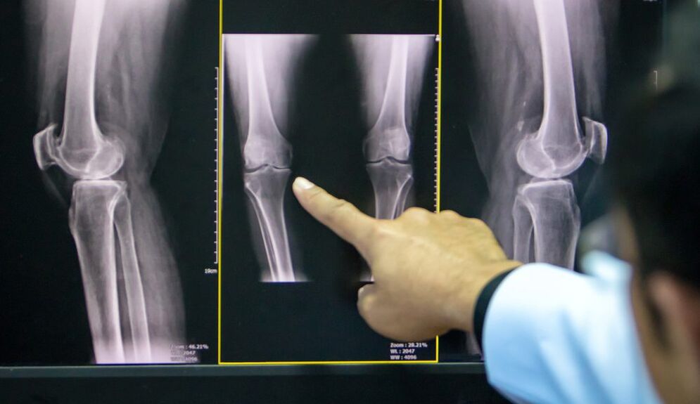
- X-ray of the knee joint in two projections (anteroposterior and lateral): the most accessible way to confirm the diagnosis in the advanced stage of pathology;
- Ultrasound: determining the presence of an effusion in the joint, measuring the thickness of the cartilage;
- analysis of synovial fluid;
- diagnostic arthroscopy (visual assessment of the cartilage) with biopsy;
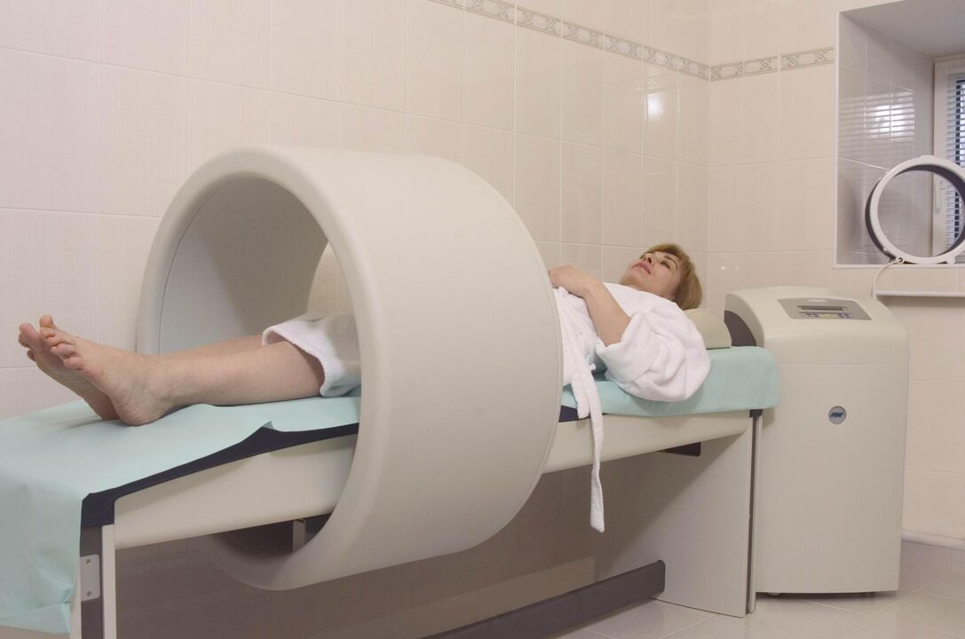
- Computed and magnetic resonance imaging (CT, MRI): the best method for diagnosing DOA in the early stages.
If the doctor has doubts about the diagnosis, it can be prescribed:
- Scintigraphy: scanning the joint after introducing a radioactive isotope;
- Thermography: study of the intensity of infrared radiation (its intensity is directly proportional to the intensity of the inflammation).
Treatment of gonarthrosis of the knee joint
The treatment regimen for arthrosis combines several approaches: non-drug methods, pharmacotherapy and surgical correction. The ratio of each method is determined individually for each patient.
Non-drug treatment
In the latest ESCEO (European Society for the Clinical Aspects of Osteoporosis and Osteoarthritis) guidelines for the treatment of knee osteoarthritis, experts place particular emphasis on patient education and lifestyle adjustments.
The patient needs:
- explain what is the essence of the disease, which is set up for long-term treatment;
- teach how to use aids (sticks, orthoses);
- prescribe a diet (for patients with a body mass index of more than 30);
- give a set of exercises to strengthen the thigh muscles and relieve the knee joint;
- Explain the importance of increased physical activity.
In the early stages of knee arthrosis, physiotherapeutic treatment methods give good results:
- Massage;
- magnetotherapy;
- UHF therapy;
- electrophoresis;
- hydrogen sulfide baths;
- paraffin applications;
- Acupuncture.
Pharmacotherapy of gonarthrosis
Drug use in DOA is aimed at relieving pain, reducing inflammation, and slowing the rate of cartilage destruction.
Symptomatic treatment:
- analgesics;
- non-steroidal anti-inflammatory substances (NSAIDs) from the group of COX-2 inhibitors in the form of tablets or suppositories;
- non-narcotic analgesics (with resistant pain syndrome).
Structure-modifying drugs (chondroprotectors):
- chondroitin sulfate;
- glucosamine sulfate.
These drugs can be taken in the form of capsules several times a year, injected intramuscularly or directly into the synovial cavity.
Local therapy includes near and intra-articular injections of glucocorticosteroids, hyaluronic acid preparations.
At stages I–II of DOA, an important place in complex therapy is the use of anti-inflammatory ointments, gels and creams based on NSAIDs. They help reduce the need for the patient to take NSAIDs by mouth, thereby reducing the risk of damage to the digestive tract.
home remedies
The use of tinctures, decoctions, extracts, local applications of medicinal plants should be considered a supportive method of treating DOA, folk remedies cannot replace the therapy prescribed by the doctor.
Plants used in arthrosis: dandelion, ginger, Jerusalem artichoke, burdock, garlic, sea buckthorn.
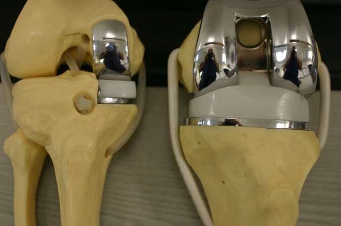
surgery
Surgical intervention may be required at all stages of gonarthrosis with insufficient effect of medical measures. Endoscopic interventions are the most common, in the most severe cases a replacement of the endoprosthesis is indicated.
Types of endoscopic interventions:
- Revision and rehabilitation of the joint: extraction of inflammatory contents from the synovial cavity, cartilage fragments;
- Plasma or laser ablation: removal of mechanical obstructions in the synovial cavity;
- chondroplasty.
A corrective periarticular osteotomy is indicated in patients with early manifestations of axial limb deformity (no more than 15 to 20%).
The purpose of the operation is to restore the normal configuration of the joint, evenly distribute the load on the joint surface and remove damaged areas. This procedure allows you to delay endoprosthetics.
Indications for replacing the affected area (or the entire joint) with an artificial one:
- DOA II-III degree;
- severe axial deformity of the limb;
- aseptic necrosis of the subchondral bone layer;
- persistent pain syndrome.
Contraindications for knee arthroplasty:
- total loss of the joint;
- unstable ligaments;
- DOA secondary to inflammatory arthritis;
- persistent flexion contracture, severe muscle weakness.
In this case, the patient undergoes arthrodesis - a comparison of the knee joint in a physiological position with the removal of the articular surfaces. This relieves pain but shortens the leg and causes secondary lesions in the contralateral knee, hip, and spine.
prevention
Prevention of premature cartilage degradation should begin in childhood.
Precautions:
- scoliosis prevention;
- Correction of flat feet (shoes with arch);
- regular physical education classes (restrict heavy sports);
- Exclusion of fixed postures during work.